Concept map synovial joint movements – Concept mapping synovial joint movements unveils a powerful tool for visualizing and analyzing the intricate dynamics of these essential body structures. Through a comprehensive examination of synovial joint types, components, and advanced techniques, this exploration delves into the multifaceted applications of concept maps in fields ranging from rehabilitation to education.
Introduction
Synovial joints, a type of freely movable joint, are characterized by their articular surfaces lined with a thin layer of hyaline cartilage and a joint cavity filled with synovial fluid. These joints facilitate a wide range of movements, making them essential for everyday activities like walking, running, and reaching.
Concept maps are valuable tools for analyzing joint movements as they provide a visual representation of the complex interactions between different anatomical structures. By mapping out the relationships between bones, muscles, ligaments, and other relevant components, we can gain a deeper understanding of how these elements work together to produce specific movements.
Key Characteristics of Synovial Joints, Concept map synovial joint movements
- Articular surfaces covered with hyaline cartilage, providing a smooth, gliding surface for movement.
- Joint cavity filled with synovial fluid, which acts as a lubricant and shock absorber.
- Surrounded by a joint capsule, a fibrous membrane that encloses the joint and provides stability.
- Reinforced by ligaments, strong bands of connective tissue that connect bones and prevent excessive movement.
Purpose of Concept Maps in Joint Movement Analysis
Concept maps facilitate a systematic approach to analyzing joint movements by:
- Visualizing the relationships between anatomical structures involved in movement.
- Identifying the primary movers and stabilizers responsible for specific movements.
- Understanding the biomechanics of joint movement, including the direction, range, and stability.
- Predicting potential movement impairments or dysfunctions based on anatomical relationships.
Types of Synovial Joints

Synovial joints, characterized by a fluid-filled synovial cavity and a lining of synovial membrane, exhibit diverse structures and functions. Based on their structural and functional characteristics, synovial joints are classified into several types, each with unique movements and capabilities.
Plane Joints
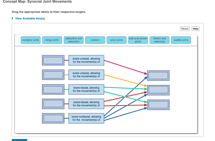
- Structure:Plane joints feature flat or slightly curved articular surfaces that allow gliding movements.
- Function:They facilitate sliding motions in various directions, such as back-and-forth or side-to-side movements.
- Examples:
- Acromioclavicular joint (between the clavicle and acromion)
- Intercarpal joints (between the wrist bones)
Hinge Joints
- Structure:Hinge joints possess cylindrical articular surfaces that permit bending and straightening motions.
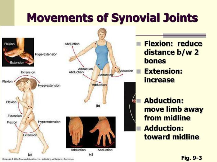
- Function:They allow for flexion (bending) and extension (straightening) movements in a single plane.
- Examples:
- Knee joint (between the femur and tibia)
- Elbow joint (between the humerus and radius)
Pivot Joints
- Structure:Pivot joints have a ring-shaped articular surface that rotates around a central axis.
- Function:They enable rotational movements around a single axis, such as turning or twisting.
- Examples:
- Atlantoaxial joint (between the first and second cervical vertebrae)
- Proximal radioulnar joint (between the radius and ulna)
Condyloid Joints
- Structure:Condyloid joints have oval-shaped articular surfaces that fit together like an egg in a spoon.
- Function:They allow for flexion, extension, abduction (moving away from the body’s midline), and adduction (moving toward the body’s midline), as well as some circumduction (circular movements).
- Examples:
- Radiocarpal joint (between the radius and carpal bones)
- Metacarpophalangeal joints (between the metacarpals and phalanges)
Saddle Joints
- Structure:Saddle joints have saddle-shaped articular surfaces that resemble a saddle.
- Function:They permit flexion, extension, abduction, adduction, and circumduction.
- Examples:
- Carpometacarpal joint of the thumb (between the trapezium and first metacarpal)
Ball-and-Socket Joints
- Structure:Ball-and-socket joints feature a ball-shaped articular surface that fits into a cup-shaped articular surface.
- Function:They provide the widest range of motion, allowing for flexion, extension, abduction, adduction, circumduction, and rotation.
- Examples:
- Hip joint (between the femur and pelvis)
- Shoulder joint (between the humerus and scapula)
Components of a Synovial Joint

Synovial joints, the most common type of joint in the body, consist of several key components that work together to facilitate movement and provide stability. These components include articular cartilage, synovial membrane, and ligaments.
Articular Cartilage
Articular cartilage is a specialized type of cartilage that covers the ends of bones at the joint surface. It is composed of a dense network of collagen fibers embedded in a matrix of proteoglycans. This structure provides the cartilage with its characteristic smoothness and resilience, allowing for frictionless movement of the joint surfaces.
Synovial Membrane
The synovial membrane is a thin layer of tissue that lines the joint cavity. It secretes synovial fluid, a viscous fluid that provides lubrication and nourishment to the joint. The synovial membrane also contains specialized cells that remove waste products and debris from the joint.
Ligaments
Ligaments are tough bands of connective tissue that connect bones to bones. They provide stability to the joint by limiting excessive movement and preventing dislocation. Ligaments are composed primarily of collagen fibers, which are arranged in a parallel fashion to provide strength and flexibility.
Together, these components work in harmony to ensure the smooth, pain-free movement of synovial joints. The articular cartilage provides a frictionless surface for movement, the synovial membrane lubricates and nourishes the joint, and the ligaments provide stability and prevent excessive movement.
Concept Mapping Joint Movements
Concept maps are a powerful tool for visually representing and analyzing joint movements. They provide a clear and concise way to see how different muscles, bones, and joints work together to produce movement.To create a concept map for a specific joint, follow these steps:
Identify the joint
The first step is to identify the joint you want to map. This could be a major joint like the knee or shoulder, or a smaller joint like the wrist or ankle.
Identify the bones involved
Once you have identified the joint, you need to identify the bones that are involved in the joint. These bones will be the nodes in your concept map.
Identify the muscles that cross the joint
The next step is to identify the muscles that cross the joint. These muscles will be the links in your concept map.
Draw the concept map
Once you have identified the bones and muscles involved in the joint, you can start to draw your concept map. The bones will be represented by circles, and the muscles will be represented by lines. The lines will connect the bones that the muscles cross.
Label the concept map
Once you have drawn the concept map, you need to label it. The labels should include the names of the bones and muscles, as well as the movements that the muscles produce.
Applications of Concept Maps: Concept Map Synovial Joint Movements

Concept maps are valuable tools that extend beyond rehabilitation and biomechanics. Their versatility allows for diverse applications across various fields, including education and beyond.
In the realm of education, concept maps serve as effective teaching and learning aids. They simplify complex concepts, enhance comprehension, and facilitate critical thinking. Students can actively engage with the material, making connections and identifying relationships between different concepts. This approach promotes a deeper understanding and retention of knowledge.
Advanced Techniques

Advanced techniques for analyzing joint movements using concept maps offer enhanced insights into complex biomechanical processes.
These techniques employ sophisticated tools and computational methods to capture and simulate joint movements with greater accuracy and detail.
Motion Capture Analysis
Motion capture analysis utilizes sensors and cameras to record the precise movements of a subject. This data is then used to create digital models of the joint and surrounding structures.
By analyzing the motion data, researchers can identify patterns, quantify joint angles, and assess the forces acting on the joint during movement.
Computer Simulations
Computer simulations leverage computational models to simulate joint movements and predict their behavior under various conditions.
These simulations incorporate factors such as muscle forces, joint geometry, and external loads to generate realistic representations of joint movement.
By manipulating simulation parameters, researchers can explore different scenarios and optimize joint design and function.
Question & Answer Hub
What are the key characteristics of synovial joints?
Synovial joints are characterized by their freely movable articular surfaces, enclosed by a joint capsule lined with synovial membrane, and filled with synovial fluid for lubrication.
How can concept maps be used to analyze joint movements?
Concept maps provide a visual representation of joint movements, allowing for the identification of patterns, relationships, and potential limitations.